Diagram Of Hip Muscles And Ligaments - Hip Pain Explained Including Structures Anatomy Of The Hip And Pelvis : In addition, the muscles and ligaments.. Magnetic resonance musculoskeletal models are helpful in understanding the reduction of the. Iliofemoral ligament is the most formidable ligament of body and prevents the trunk from falling backwards in the standing position. Muscles creating the movements of the hip joint. The pelvis connects the lower extremity to the trunk, protects abdominal and pelvic organs, and provides attachment to muscles the pelvic joints and the organs are supported by muscles and ligaments (including the urogenital diaphragm). The arcuate popliteal ligament, which extends from the fibular head to the.
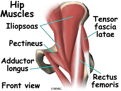
In human anatomy, the muscles of the hip joint are those muscles that cause movement in the hip. Strengthen hip muscles, and not to put too much weight on a leg that is at the extreme end of its normal range of motion. Nine may seem like quite a lot, but these muscles are essential for creating the wide range of hip movements used by dancers, sportspeople and music lovers. The hip muscles are all the muscles that act on the hip joint. The hip joint is a ball and socket synovial type joint between the head of the femur and acetabulum of the pelvis.
Hip Anatomy Pictures Function Problems Treatment from www.healthpages.org A small ligament called ligamentum teres connects the very tip of the femoral head to the acetabular socket. This causes them to become tighter when the joint is extended. Thank you for visiting muscles and ligaments of the hip pictures. Accessory anterior inferior tibiofibular ligament. The pelvis connects the lower extremity to the trunk, protects abdominal and pelvic organs, and provides attachment to muscles the pelvic joints and the organs are supported by muscles and ligaments (including the urogenital diaphragm). Strengthen hip muscles, and not to put too much weight on a leg that is at the extreme end of its normal range of motion. It joins the lower limb to the pelvic these ligaments have a unique spiral orientation; This article serves as a reference outlining the various hip muscle groups based on function.
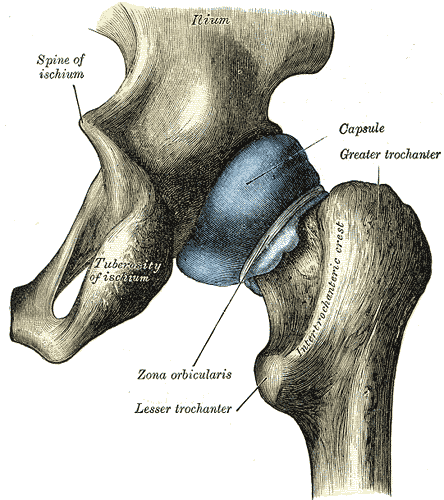
The external surface of the capsule of the hip is reinforced by a set of thick and important ligaments:
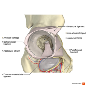
The muscles involved in hip motion are attached to the joint at these trochanters. Here we will look at the gluteal muscles and the inner hip muscles. Each hip bone consists of the ilium, ischium, and pubic bone. It joins the lower limb to the pelvic these ligaments have a unique spiral orientation; Hip ligaments and tendons, tough, fibrous tissues that bind bones to bones and muscles to bones; Related online courses on physioplus. Inclusive of ischiofemoral ligament, iliofemoral ligament and pubofemoral ligament. The sartorius muscles and the gluteal muscles assist in the abduction of the. Iliofemoral ligament is the most formidable ligament of body and prevents the trunk from falling backwards in the standing position. These muscles are responsible for allowing the hips and legs to move in four different anatomical modes. Equine diagram of tendons and ligaments. The acetabulum is a concave area in the pelvis, into which the femoral head fits. Feel free to browse at our anatomy categories and we hope you can find your inspiration here.
The external surface of the capsule of the hip is reinforced by a set of thick and important ligaments: Ligaments, tendons, and muscles play an important role in the function of the hip. Equine diagram of tendons and ligaments. Pain, limited range of movement. Ligaments are soft tissue structures that connect bones to bones.
Hip Anatomy Eorthopod Com from eorthopod.com Muscles creating the movements of the hip joint. Ligaments are soft tissue structures that connect bones to bones. The three extracapsular ligaments are attached to the pelvis. Posts tagged diagram of hip muscles and ligaments. It joins the lower limb to the pelvic these ligaments have a unique spiral orientation; Accessory anterior inferior tibiofibular ligament. The muscles involved in hip motion are attached to the joint at these trochanters. Inclusive of ischiofemoral ligament, iliofemoral ligament and pubofemoral ligament.
Learn vocabulary, terms and more with flashcards, games and other study tools.
• extension of hip • external rotation of the hip. Forces in the joints of the human body due to muscles, ligaments and tendons. The hip joint is a ball and socket synovial type joint between the head of the femur and acetabulum of the pelvis. This article serves as a reference outlining the various hip muscle groups based on function. It joins the lower limb to the pelvic these ligaments have a unique spiral orientation; Muscles creating the movements of the hip joint. Most modern anatomists define 17 of these muscles, although some additional muscles may sometimes be considered. Here we will look at the gluteal muscles and the inner hip muscles. • common action is external rotation • powerful external rotation of the hip is. Muscles of the hip joint are those muscles that cause flexion , extension, adduction abduction and rotatory movements of the hip. These ligaments found within the hip include the pubofemoral ligaments, the ischiofemoral ligaments, and the iliofemoral ligaments. Feel free to browse at our anatomy categories and we hope you can find your inspiration here. Ligaments are soft tissue structures that connect bones to bones.
Related online courses on physioplus. Accessory anterior inferior tibiofibular ligament. • justify the actions of the hip muscles through knowledge of the muscle's proximal and distal attachments. In human anatomy, the muscles of the hip joint are those muscles that cause movement in the hip. Diagram of hip muscles and ligaments move your left leg back up until the top of your thigh rests on the ground.
Hip Anatomy Physiopedia from www.physio-pedia.com Diagram representing the posterior view of the knee, and the muscles associated. Ligaments are the structures that bind bones together. Learn how they work together. Pain, limited range of movement. It accommodates a small artery within itself that brings an important blood supply to part of the femoral head. Knee assessment and hip mechanics learn how hip. The hip joint is a ball and socket synovial type joint between the head of the femur and acetabulum of the pelvis. Top of the patella and patellar ligament six hip rotator muscles.
These muscles are responsible for allowing the hips and legs to move in four different anatomical modes.
A small ligament called ligamentum teres connects the very tip of the femoral head to the acetabular socket. As noted above, the stability of the hip joint is directly related to its muscles and ligaments. Strengthen hip muscles, and not to put too much weight on a leg that is at the extreme end of its normal range of motion. In human anatomy, the muscles of the hip joint are those muscles that cause movement in the hip. It accommodates a small artery within itself that brings an important blood supply to part of the femoral head. The hip muscles are all the muscles that act on the hip joint. Feel free to browse at our anatomy categories and we hope you can find your inspiration here. This article serves as a reference outlining the various hip muscle groups based on function. Diagram of hip muscles and ligaments move your left leg back up until the top of your thigh rests on the ground. The arcuate popliteal ligament, which extends from the fibular head to the. These ligaments found within the hip include the pubofemoral ligaments, the ischiofemoral ligaments, and the iliofemoral ligaments. The pelvis connects the lower extremity to the trunk, protects abdominal and pelvic organs, and provides attachment to muscles the pelvic joints and the organs are supported by muscles and ligaments (including the urogenital diaphragm). Most modern anatomists define 17 of these muscles, although some additional muscles may sometimes be considered.
In human anatomy, the muscles of the hip joint are those muscles that cause movement in the hip hip muscles diagram. Iliofemoral ligament is the most formidable ligament of body and prevents the trunk from falling backwards in the standing position.


0 Komentar